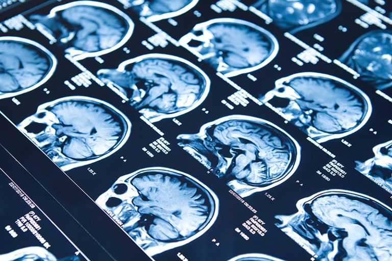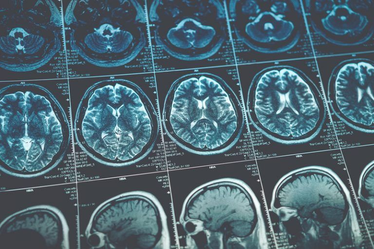
Neuroimaging
We have been representing individuals with brain injuries for over 30 years. We represent individuals with brain injuries that are mild, moderate, and severe. When establishing a mild traumatic brain injury (i.e. a concussion), there is an art to being able to prove that injury to an insurance adjuster, defense counsel, judge, jury, etc. This is largely because brain injuries are “invisible” – they are unlike other injuries, such as broken bones that you can easily identify on an x-ray. However, in certain cases, the invisible injury can now become visible with the use special brain injury trauma protocols for neuroimaging.
When we first began representing individuals with brain injuries, options for proving and establishing a mild traumatic brain injury were limited. We did not have the ability to go get an image of the brain to show the areas of damage caused by a mild traumatic brain injury. The neuroimaging that was available would typically only show the severe TBIs, such as bleeding and hemorrhaging in the brain or fractures in the skull. Rather, one of the only ways to “objectively” establish a mild traumatic brain injury was through a clinical evaluation process that included neuropsychology. The objectivity was the clinical similarities with other brain-injured individuals and neuropsychology. However, many people would argue that neuropsychology is not objective and is subject to great variations in interpretation.
Progression of Neuroradiology
Fortunately, over the past 15 years, the progression and growth of neuroradiology has been astounding in the area of brain injury. The old saying, “a picture is worth a thousand words,” is very accurate when attempting to prove a brain injury. The larger MRI magnets that are now available (3 Tesla, 5 Tesla, and 7 Tesla magnets) have allowed us to see so much more detail. There are additionally other modalities of imaging that can identify brain damage. Because of these advances we are able to see objective evidence of axonal shearing, microscopic bleeds, lack of water flow due to brain damage, and atrophy due to trauma – things we could not identify 15 years ago.
Utilizing the newer MRI technology has been earth-shattering in establishing brain injuries objectively. As a result, the ability to prove brain injuries in a legal setting has been made clearer, more objective, and easier for a lay person to understand and to visualize. When we have assembled focus groups for brain injury cases, the jurors continually anchor their decisions to the neuroradiology results. These results make a major difference in proving the legal case and in the size of the recovery.
Neuroimaging, however, requires doctors to clinically correlate these objective imaging findings to the symptoms of a brain-injured person.
An example of clinical correlation is as follows:
Patient X was a healthy individual with no prior concussions or symptoms. Patient X was in a car accident and sustained a concussion. The concussion does not resolve and develops into postconcussion syndrome with ongoing symptoms of depression, emotional instability, changes in behavior, reduced creativity, and impaired judgment (all frontal lobe injury symptoms). If imaging shows that there is damage to the frontal lobe of the brain, there is now clinical correlation between the symptoms the patient is suffering from and the damage that is shown on the brain images. Thus, the doctor would opine that in all probability the damage on the brain images came from the car accident and it is THE CAUSE of Patient X’s symptoms.
To tie these findings to the patient’s clinical picture is invaluable in the clinical, as well as legal, settings.
Better Care
In addition to helping prove the brain injury, the objectivity and the ability to compare those findings with the rest of the clinical picture has been incredibly helpful in the area of providing individuals with more effective care. Understanding multiple layers of objective proof is indispensable for providing care providers with more information in order for them to offer more effective and efficient care through occupational therapy, speech therapy, neurological physical therapy, and vocational rehabilitation.
Detailed neuroimaging is not available everywhere. For years we have asked our clients to go to New York, Detroit, and Las Vegas to obtain specialized brain trauma protocol MRIs. We did not have the ability to get the trauma protocol done locally and have it read by the most experienced doctors in the field. The trauma protocol includes thinner slices, diffusion tensor imaging, neuroquant (brain atrophy), susceptibility-weighted imaging, gradient echo, T2, and flair sequences. This can all now be done in the Denver metropolitan area. The neuroradiological read is done by a high level neuroradiologist in Las Vegas who is willing to testify as to his findings and work with the local providers.
Brain Injury Representation
We pride ourselves in providing you with the highest level of representation to resolve your claims for the highest recovery possible. Our experience and knowledge in brain injury not only allows us to know and gather the information necessary to prove your claim, but also allows us to understand your injury and what you are going through and to be able to put you in contact with quality care providers. Have you suffered a brain injury through the fault of another person or entity? Please contact us for more information on how we can help you with your brain injury case.